|
|
|
|
|
|
| |
|
|
| |
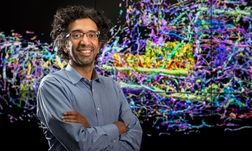
Neurobiologist Bobby Kasthuri wants to map the neural universe. |
|
| |
|
|
|
| |
|
|
| |
|
|
| |
| |
|
|
| |
(Photography by Mark Lopez/Argonne National Laboratory) |
|
| |
|
|
|
|
| |
|
|
|
| |
|
|
| |
To learn how a machine works, look inside at how it's wired. |
|
| |
|
|
|
| |
|
|
| |
UChicago and Argonne National Laboratory scientists are doing just that--combining new techniques in microscopy, neurobiology, and computing--to reveal the brain's inner mechanisms in unprecedented detail. |
|
| |
|
|
|
| |
|
|
| |
The brain is like a computer, operating on electrical impulses and chemical signals sent between cells. Though microscopic, brain wiring is theoretically possible to diagram, as has been done for simple organisms. |
|
| |
|
|
|
| |
|
|
| |
But the human brain is not so simple. It contains nearly 100 billion neurons, scientists estimate, each making tens of thousands of contacts with other cells. A complete map of these connections--sometimes called the connectome--would be the largest dataset ever created. |
|
| |
|
|
|
| |
|
|
| |
"It's a huge theory of neuroscience that all of our behaviors, all of our pathologies, all of our illnesses, all of the learning that we do, is all due to changes in the connections between brain cells," says UChicago and Argonne neurobiologist Narayanan "Bobby" Kasthuri. He's working to test this theory using powerful microscopes and supercomputers at Argonne. |
|
| |
|
|
|
| |
|
|
| |
|
|
| |
| |
|
|
| |
Bobby Kasthuri, hired in 2015 as Argonne's first neuroscientist, explains the connectome at five levels of difficulty, from simple enough for a five-year-old to complex enough for a connectome investor. |
|
| |
|
|
|
|
| |
|
|
|
| |
|
|
| |
Viewing synapses requires an electron microscope to image a slice of brain thousands of times thinner than a sheet of paper. Kasthuri has developed a method, using a diamond knife with an edge only five atoms thick, to cut 50-nanometer-thin slices of human or animal brain. The slices then float through water to a conveyer belt, which takes each one beneath the gaze of an electron microscope. |
|
| |
|
|
|
| |
|
|
| |
Next, the images are analyzed by the Theta supercomputer, typically used to process data from CERN's Large Hadron Collider or run universe expansion models in the search for dark matter. But mapping an entire human brain, which would require 300,000 of these images, and reconstructing the whole organ in 3D, is beyond the capabilities of even this world-class machine. |
|
| |
|
|
|
| |
|
|
| |
So Kasthuri is starting (relatively) small. Rather than build a complete map of the human brain, he is focusing first on comparing connection patterns between brains young and old, animal and human, typical and those of people with mental disorders--which may one day influence clinical treatment. |
|
| |
|
|
|
| |
|
|
| |
But applying connectome research to medicine is a long road. The near-term benefits of brain mapping will be to equip scientists studying elemental links between brain and behavior with a deeper understanding of the organ's complex mechanical structure. |
|
| |
|
|
|
| |
|
|
| |
Learn more from Kasthuri's Big Brains podcast interview, where he discusses AI's ability to bluff, the physical basis of mental illness, and the advantages of baby brains.--Rob Mitchum |
|
| |
|
|
|
|
|
| |
|
|
| |
Five more imaging innovations |
|
| |
|
|
|
| |
|
|
| |
|
|
|
| If you compare brain imaging to Google Maps, functional MRI gives you the satellite view and microscopy gives you the street view. Now, powerful x-rays can give you the navigational map view, too. |
|
|
|
| |
|
|
|
|
|
|
|
|
|
| |
|
|
| |
|
|
|
| MBL researchers investigate regeneration using high-resolution microscopy and time-lapse video of Earth's largest single-celled organism. |
|
|
|
| |
|
|
|
|
|
|
|
|
|
|
|
|
|
|
|
|
|
| |
|
|
| |
|
|
| |
| |
|
|
| |
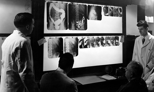
Paul C. Hodges (seated at right), professor and founding chair of the UChicago Department of Radiology, joined the University in 1927, the year it matriculated its first class of medical students. He views x-rays with colleagues in this 1950 photo. (Photography by John Kaspar. UChicago Photographic Archive, apf1-02829, University of Chicago Library) |
|
| |
|
|
|
|
| |
|
|
|
| |
|
|
| |
Born in 1893, before the discovery of x-rays, Paul C. Hodges began his work in radiology at age 14, apprenticing at the Ashland, Wisconsin, hospital where his uncle worked and learning to prepare chemical solutions used for "x-ray photography." |
|
| |
|
|
|
| |
|
|
| |
He served as a radiologist through World War I and then at the Peking Union Medical College in China before traveling to UChicago for a yearlong sabbatical that turned into a 31-year career, during which he founded the radiology department. |
|
| |
|
|
|
| |
|
|
| |
To aid his work improving subspecialty applications, film development facilities, and novel hardware, he established a dedicated machine shop--uncommon in hospitals at the time. |
|
| |
|
|
|
| |
|
|
| |
Dissatisfied with manufactured equipment, Hodges worked with engineers and machinists to design and build his own specialized x-ray machines and tables as well as meters to measure and timers to control radiation exposure. |
|
| |
|
|
|
| |
|
|
| |
He also partnered with the 3M Company to improve compounds for attaching jacks to the ends of high-voltage cables, the Eastman Kodak Company to obtain its brand-new automated x-ray film processor, and Nestlé to create chocolate-flavored barium mixtures for gastrointestinal exams. |
|
| |
|
|
|
| |
|
|
| |
While advancing the field, Hodges's department treated several hundred thousand patients before he retired in 1958. |
|
| |
|
|
|
|
|
|
|
|
|
| |
|
|
| |
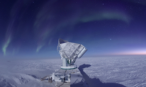
Photogenic:
South Pole Telescope points newest camera at big bang's light.
|
| |
|
|
|
|
| |
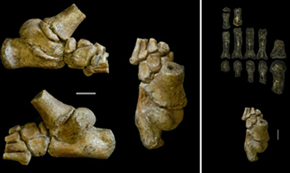
Limber:
Ancient toddlers climbed trees with the help of a highly mobile big toe.
|
| |
|
|
|
|
|
|
|
|
|
|
| |
|
|
| |
| |
|
|
| |
Good things come to those who share! Forward µChicago to a friend and be entered to win a prize. Congratulations to last month's contest winner, James Dricker, AB'70.
Subscribe to receive µChicago monthly. |
|
| |
|
|
|
|
| |
|
|
|